Microscopic Investigation
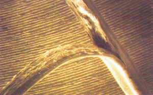
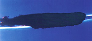
The hair shaft provides important diagnostic clues to such disorders as congenital defects, external damage to hair, general illness, fungal infection of the hair and scalp and developing areas of baldness.
The hair passes through three phases, anagen, catagen and telogen, with each phase dramatic cell changes take place. Under a microscope the living hair bulb represents the bodys health and stability. The hair shaft is like a diary of what has happened over a period of time. Many body disorders , acute or chronic , local or systemic, will be reflected in the bulb and eventually in the shaft. Thus the cells of the hair provide an exceptionally sensitive index to the constantly changing internal environments as well as a reflection of events occuring in the body’s exchange with its external world.
Some conditions such as protein malnutrition or the body’s response to chemotherapeutic drugs prescribed for cancer, produce remarkably specific changes in the size, structural arrangement even the pigmentation of the hair bulb.
A microscopic investigation can;
Assess structural damage.
Establish the rate of hair loss.
Determine the severity of hair loss.
Confirm the presence of lice or fungus.
Identify genetic problems.


These photos are the copyright of the International Association of Trichologists and David Salinger and are reproduced with their permission.
